

Concerns regarding Transfusions of Blood Products Derived from GeneticVaccine Recipients and Proposals forSpecific Measures
Preprints.org Review: Not peer-reviewed version
Jun Ueda * , Hideyuki Motohashi , Yuriko Hirai , Kenji Yamamoto , Yasufumi Murakami , Masanori Fukushima , Akinori Fujisawa *
Posted Date: 15 March 2024
doi: 10.20944/preprints202403.0881.v1
Review
Concerns regarding Transfusions of Blood Products
Derived from Genetic Vaccine Recipients and
Proposals for Specific Measures
Jun Ueda 1,*, Hideyuki Motohashi 2, Yuriko Hirai 3, Kenji Yamamoto 4, Yasufumi Murakami 5,
Masanori Fukushima 6 and Akinori Fujisawa 7,*
1 Department of Advanced Medical Science, Asahikawa Medical University, Asahikawa 078-8510,
Hokkaido, Japan; junueda@asahikawa-med.ac.jp
2 Pre-Clinical Research Center, Tokyo Medical University Hospital, 6-7-1 Nishi-Shinjuku, Shinjuku-ku,
Tokyo 160-0023, Tokyo, Japan; moto@tokyo-med.ac.jp
3 MCL Corporation, Jimukino-Ueda bldg. 603, 21 Sakaimachi Gojo-Takakurakado, Shimogyo-Ku, Kyoto 600-
8191, Kyoto, Japan; hirai@mcl-corp.jp
4 Department of Cardiovascular Surgery, Center of Varicose Veins, Okamura Memorial Hospital, 293-1
Kakita Shimizu-cho, Sunto-gun, Shizuoka 411-0904, Japan; yamamoto@okamura.or.jp
5 Department of Biological Science and Technology, Faculty of Advanced Engineering, Tokyo University of
Science, 6-3-1 Niijuku, Katsushika-ku, Tokyo 125-8585, Japan; yasufumi@rs.tus.ac.jp
6 Foundation of Learning Health Society Institute, Nagoya 450-0003, Aichi, Japan; mfukushima@imrd.jp
7 Kokoro Medical Corporation, Honbetsu Cardiovascular Medicine Clinic, Honbetsu 089-3314, Hokkaido,
Japan; fujisawa.peace@mac.com
* Correspondence: junueda@asahikawa-med.ac.jp (J.U.); fujisawa.peace@mac.com (A.F.);
Tel.: +81-166-68-2385 (J.U.); +81-156-22-8888 (A.F.)
Abstract: The coronavirus pandemic was declared by the World Health Organization (WHO) in
2020, and a global genetic vaccination program has been rapidly implemented as a fundamental
solution. However, many countries around the world have reported that so-called genetic vaccines,
such as those using modified mRNA encoding the spike protein and lipid nanoparticles as the drug
delivery system, have resulted in post-vaccination thrombosis and subsequent cardiovascular
damage, as well as a wide variety of diseases involving all organs and systems, including the
nervous system. In this article, based on these circumstances and the volume of evidence that has
recently come to light, we call the attention of medical professionals to the various risks associated
with blood transfusions using blood products derived from people who have suffered from long
COVID and from genetic vaccine recipients, including those who have received mRNA vaccines,
and we make proposals regarding specific tests, testing methods, and regulations to deal with these
risks. We expect that this proposal will serve as a basis for discussion on how to address postvaccination
syndrome and its consequences following these genetic vaccination programs.
Keywords: COVID-19 vaccine; genetic vaccine; blood product; blood transfusion; spike protein;
post-vaccination syndrome; harm–benefit assessment; prion; spikeopathy; inspection standard;
diagnostic criteria
1. Introduction
On March 11, 2020, the coronavirus pandemic was declared by the Director-General of the World
Health Organization (WHO) [1], and countries actively implemented classical public health
measures, including quarantine, isolation, disinfection, and lockdowns. However, hopes for a vaccine
grew as the general consensus was that rapid herd immunity was the best solution to overcome the
pandemic. Since 2021, as a means to combat SARS-CoV-2 infection, several global pharmaceutical
companies including Pfizer-BioNTech, Moderna, and AstraZeneca have developed various genetic
vaccines that use the spike protein of the Wuhan strain of SARS-CoV-2 as an antigen, and rapid
vaccination has been promoted on a global scale [2,3]. During this period, virological studies of SARSCoV-
2 have been intensively conducted, and the pathogenic mechanism of this virus has been
elucidated in detail [4,5]. In brief, the key pathogenic processes include the binding of the spike
protein of SARS-CoV-2 to the angiotensin-converting enzyme 2 (ACE2) receptor on vascular
endothelial cells, allowing viral entry and amplification [6]; the triggering of red blood cell and
platelet aggregation by the spike protein [7–11]; and the formation of microthrombi [12,13].
However, it has been reported from various countries around the world that genetic vaccines
such as mRNA vaccines encoding spike proteins have also caused a wide variety of diseases in all
organs and systems, including the nervous system, in addition to thrombosis and resulting
cardiovascular disorders in vaccine recipients [14–21]. This is because when the foreign gene was
introduced into autologous cells using gene-transfer capable lipid nanoparticles (LNPs) or other
means, the spike proteins produced from the mRNA or DNA introduced via the gene vaccine
induced thrombosis in the vaccine recipient. While evidence for specific problems has been reported
individually, Parry et al. have proposed the theory of spikeopathy (spike disease) as a hypothesis
that synthesizes all of the evidence for this problem [22]. Furthermore, there are two general
mechanisms by which a modified gene introduced into the body by genetic vaccination and some of
the antigens produced because of the expression of that gene can be transmitted throughout the body.
First, LNPs encapsulating mRNA can spread through the body via the bloodstream from the injection
site. It has already been shown that LNPs have a tendency to accumulate in specific organs, such as
the liver, spleen, ovaries, testes, and bone marrow [22,23]. The other is the release of
pseudouridinated mRNA molecules and synthesized spike proteins as extracellular vesicles, or
exosomes, from cells that have incorporated LNPs. These exosomes are transported in the circulation
throughout the body to reach various organs [24–27]. And it has already been proven that spike
proteins produced by cells that have taken up the modified gene travel throughout the body in the
bloodstream [28,29]. Thus, it must be emphasized that the transport, distribution, and expression of
the components of the genetic vaccine beyond the administration site to organs and tissues of the
whole body after vaccination involve the risk of inducing various conditions.
Although the Director-General of the WHO declared the end of the COVID-19 public health
emergency on May 5, 2023, post-vaccination syndrome (PVS), caused by genetic vaccines that have
been promoted worldwide and have been given to billions of people, has become a major global
problem [19,21,27,30] requiring a reasonable harm–benefit assessment of the global use of genetic
vaccines [27,31–33]. Since the beginning of the coronavirus pandemic and genetic vaccination, there
has been much debate about the safety of blood products and their use in transfusions [34–39].
However, because the pathology of SARS-CoV-2 was not fully understood at the beginning, there
was no specific discussion based on data or analysis of what was a problem and what could be a risk;
only concerns were expressed, and no clear conclusions or policies were drawn. For example, Jacobs
et al. argued that there was no requirement to collect or share the genetic vaccination status of blood
donors and that hospitals were not required to inform patients about the genetic vaccination status
of blood donors [37], because there were no reports of health issues from genetic vaccination in 2021.
However, this argument was not based on data. Contrary to initial expectations, it was found that
genes and proteins from genetic vaccines persist in the blood of vaccine recipients for prolonged
periods of time [22,28,40–44], and a variety of adverse events resulting from genetic vaccines are now
being reported worldwide. Roubinian et al. reported that transfusions of plasma and platelet blood
components collected before and after COVID-19 vaccination were not associated with increased
adverse outcomes in transfusion recipients who did not develop COVID-19 [39]. However, they
evaluated only plasma and platelet preparations, not red blood cell or whole blood preparations. The
long-term effects remain unclear, as the study only followed up recipients to the point of 30-day
readmission rates.
Considering the current situation and the volume of evidence that has recently come to light,
the purpose of this article is to raise awareness among relevant parties and point toward future
directions by making specific recommendations regarding the use of blood products derived from
genetic vaccine recipients, including those who have received mRNA vaccines. To be more precise,
genetic vaccines are the equivalent of biomedicine (i.e. immune therapeutics) rather than
conventional vaccines in terms of their mechanism of action [45,46]. The various genetic vaccines now
treated as vaccines should originally have been treated as biomedicine, but because they were
classified as vaccines, huge numbers of people were inoculated with them [2,3]. As a result, extensive
areas of medicine are now beginning to be affected because most of the population in many countries
has been vaccinated [19,21,27,30,47]. This has never happened before in the history of biomedicine,
and consequently, it is highly suspected that blood products for transfusion have been affected by
these so-called genetic vaccines. Therefore, this review was prepared to examine the risks of blood
transfusions at the current stage when genetic vaccines are administered in large quantities. The
vaccine recipients described in this proposal are limited to genetic vaccine recipients.
2. Overview of Cases of Blood Abnormalities after Genetic Vaccination
A wide variety of diseases related to blood and blood vessels, such as thrombosis, have
developed after genetic vaccination, including with mRNA vaccines, and many cases of serious
health injuries have been reported. For example, a PubMed search on diseases such as
thrombocytopenia, thrombotic disorders with thrombocytopenia, deep vein thrombosis,
thrombocytopenic purpura, cutaneous vasculitis, and sinus thrombosis combined with the essential
keywords “COVID-19 vaccine” and “side effects” yielded several hundred articles in only about two
years since the rollout of genetic vaccines [14,17,20,21,48]. In addition to abnormally shaped red blood
cells, amorphous material has been found floating in the blood of mRNA-vaccinated individuals
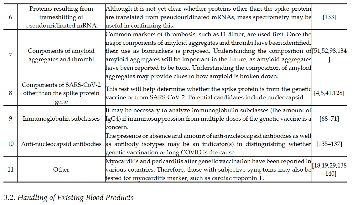
under microscopic observation, some of which has shown grossly abnormal findings (Table 1, point
5) [7–10,49]. Recent studies have also reported that the spike protein has amyloidogenic potential [50–
54], is neurotoxic [55–57], and can cross the blood–brain barrier [58–60]. Thus, there is no longer any
doubt that the spike protein used as an antigen in genetic vaccines is itself toxic [22,61,62].
In addition to thrombosis, individuals who have received multiple doses of a genetic vaccine
may have multiple exposures to the same antigen within a brief period, thereby being imprinted with
a preferential immune response to that antigen [63,64]. This phenomenon, called original antigenic
sin or immune imprinting, has caused COVID-19 vaccine recipients to become more susceptible to
contracting COVID-19 [65]. In addition, antibody-dependent enhancement of infection is also known;
antibodies produced by vaccination may rather promote viral infection and symptoms [66,67]. On
the other hand, it has also been suggested that repeated administration of genetic vaccines may result
in immune tolerance because of a class switch to non-inflammatory immunoglobulin G4 (IgG4) [68–
71], whereby the immune system of the recipient does not mount an excessive response such as
cytokine storm [27,72], and case reports of IgG4-related disease have begun to appear [73–75]. This
raises concern that alterations in immune function due to immune imprinting and immunoglobulin
class switching to IgG4 may also occur in genetic vaccine recipients. This may increase the risk of
serious illness due to opportunistic infections or pathogenic viruses that would not normally be a
problem if the immune system were normal [76–82]. For example, cases of suspected viremia have
been reported [82]. Therefore, from the perspective of traditional containment of infectious diseases,
greater caution is required in the collection of blood from genetic vaccine recipients and the
subsequent handling of blood products, as well as during solid organ transplantation and even
surgical procedures [83–87] in order to avoid the risk of accidental blood-borne infection (Table 1,
point 3) [84–87]. The phenomenon of immune imprinting can occur even when spike protein is not
used as an antigen or when another antigen is used (e.g. inactivated influenza vaccine) [88]. However,
compared to conventional inactivated vaccines, genetic vaccines, which produce an antigen within
the body, are expected to prolong the period of exposure to the same antigen, and as a result, the risk
of immune imprinting may be higher than with conventional vaccines. It is not actually known how
long the vaccine components remain in the body after a person has received a genetic vaccine
[22,40,43], but it is expected that they will remain in the body for a longer period than originally
thought, in part because spike protein has been detected in the bodies of people several months after
vaccination (Table 1, point 1) [22,28,41,42]. In addition, since long-term exposure to a specific identical
antigen (in this case, spike protein) causes immunoglobulins to become IgG4 [68,70] and some of the
B cells that produce them are likely to differentiate into memory B cells that survive in the body for
a sustained period [70,89], the immune dysfunction of genetic vaccine recipients is expected to be
prolonged (Table 1, point 3 & 6). More details on these points are expected to be revealed in the future.
In summary, there is an undeniable risk that patients may experience some problems if they
receive blood products derived from blood collected in, at least, a brief deferral period after genetic
vaccination. Although it is unknown at present whether secondary damage is caused by transfusion
of blood products derived from genetic vaccine recipients, it is necessary for medical institutions and
administrative organizations to respond and investigate cooperatively, keeping various possibilities
in mind, because mechanisms such as the toxicity of the spike protein itself and the effects of LNPs
and modified mRNA on the immune response have not been fully elucidated and are still under
study. It should be emphasized that a significant proportion of the COVID-19 PVS in mRNA vaccine
recipients is due to toxic spike proteins, and the inclusion of structures in the receptor-binding
domain within these proteins that may induce prion disease is particularly alarming, as Seneff et al.
and Perez et al. have warned [50,90–96]. Furthermore, it has been shown that prion similarity in the
receptor-binding domain exists not only in the spike protein of the Wuhan strain, which is still used
as an antigen in genetic vaccines, but also in the spike protein of variants of SARS-CoV-2, such as the
Delta strain, with the exception of the Omicron strain [93,97]. Whether we should be uniformly
vigilant for the spike protein of the coronavirus or just the spike protein of certain variants, such as
the Wuhan strain, awaits further analysis.

3. Specific Proposals for Blood Sampling and Blood Products from Vaccine Recipients
In the previous section, we discussed a variety of blood-related abnormalities that have occurred
following genetic vaccination. In this section, we provide specific proposals on how to respond to
these circumstances. Because blood contamination affects so many areas of health care, it is especially
important to anticipate the worst [95,96,108–110] and to plan and act from the start to ensure that
there are no lapses or omissions.
3.1. Additional Requirements for Blood Collection (Donation)
Currently, in Japan, the Japanese Red Cross Society (https://www.jrc.or.jp/english/) plays a
central role in blood collection activities, and its blood products are used for blood transfusions and
other purposes. The Japanese Red Cross Society has a rule that blood can be collected from genetic
vaccine recipients after a deferral period (48 hours for mRNA vaccine recipients and 6 weeks for
AstraZeneca DNA vaccine recipients), but the data and rationale for the rule have not been specified.
As with infections such as human immunodeficiency virus (HIV) and prion diseases, a history of
genetic vaccination (DNA and/or mRNA type), including timing and number of doses, should be
obtained by interview, and kept in the official record when blood is collected (Figure 1, Table 2).
Additional caution is needed, particularly if not many days have passed since the genetic vaccine
was administered, because LNPs [23,101–103] and spike protein mRNA, which can induce
inflammation, may remain in the blood (Table 1, point 4) [22,40,43,44]. If certain events such as
anaphylactic shock occur immediately after genetic vaccination, the effects of LNPs should also be
suspected [100]. It has also been reported that negatively charged LNPs themselves interact with
fibrinogen to form thrombi [99]. Therefore, the presence of LNPs may itself be a factor in the need for
caution with transfusion products.
On the other hand, even if a person has not received a genetic vaccine, if they have had long
COVID, it is possible that the spike protein remains in their body, and thus it would be better to keep
an official record of whether they have long COVID or not [51,111–113]. As the degradation rates of
pseudouridinated mRNA and spike protein in the body are unknown at present, blood products
derived from genetic vaccine recipients should be used with extreme caution, being conscious of the
cases of AIDS, bovine spongiform encephalopathy (BSE), and variant Creutzfeldt-Jakob disease
(vCJD) caused by the use of contaminated blood products in the past [110,114–121].

At present, the genetic vaccination status of blood donors is not confirmed or controlled by
organizations including medical institutions, and the use of blood collected from these donors for
transfusions may pose risks to patients. Therefore, when blood products derived from gene vaccine
recipients are used, it is necessary to confirm the presence or absence of spike protein or modified
mRNA as in other tests for pathogens (Figure 1, Table 2). These should be quantified by an
immunochemical enzyme-linked immunosorbent assay (ELISA), by immunophenotyping, by direct
mass spectrometry of the protein itself, by an exosome-based liquid biopsy as used in cancer
screening, or by PCR [28,29,122–128]. For protein assays, as it may take time to generate a goodquality
anti-spike protein antibody or a positive control for a recombinant spike protein to be
compared with, and to sort and distribute them to each laboratory, we suggest that mass
spectrometry be used as an initial step to identify and quantify the spike protein itself in blood
[28,125]. In parallel with this, an analysis of the components of the spike protein-induced amyloid
material will be needed [51,98]. Once the components of amyloid aggregates are identified, they can
be used as biomarkers in the future. Exosome analysis will also be useful as a test as it has already
been shown that spike proteins and their genes are transported in the circulation around the body by
exosomes [24–27].
If the blood product is found to contain the spike protein or a modified gene derived from the
genetic vaccine, it is essential to remove them. However, there is currently no reliable way to do so.
As noted above, the prion-like structure within the spike protein molecule [91,95,96] suggests that
this molecule may be a persistent, sparingly soluble, heat-resistant, and radiation-resistant protein
[141,142]. The prion protein can be inactivated by thiocyanate, hydroxide, and hypochlorite [143–
145], but it is not yet known whether these can be applied to the spike protein and the resulting
amyloid materials. Therefore, as there is no way to reliably remove the pathogenic protein or mRNA,
we suggest that all such blood products be discarded until a definitive solution is found. Discarding
blood products prepared from blood collected from many dedicated blood donors can be very
painful, but it is necessary because the spike protein itself has been shown to induce thrombosis and
similar diseases. However, some medical facilities may have difficulty disposing of blood products
immediately, in which case it is essential to add the possibility of contamination with spike protein
or other foreign substances to the transfusion consent form and to fully explain this to the patient. In
any case, to prevent and reduce medical accidents caused by contaminated blood, it is imperative to
underscore the importance of confirming the history and frequency of genetic vaccination at the time
of blood collection and this information should be documented as an official record, managed and
stored by both medical and governmental organizations (see Figure 1, Table 2).
3.3. The Need for Regular Checkups and Cohort Studies to Gain a Complete Picture of Blood Contamination
As the residual status of spike protein or modified gene fragments derived from genetic vaccines
is currently unknown, it will be necessary in the future to include measurement of these amounts in
routine health checkups. It is also necessary to include a section in the routine medical checkup
questionnaire to check genetic vaccination status and the number of vaccinations to obtain an overall
picture of the residual status of spike proteins in the blood. This is because a variety of conditions
following genetic vaccination involve thrombosis and immunological conditions
[12,14,16,17,21,22,68,70]. Therefore, abnormalities in blood components related to these events should
also be analyzed.
On the other hand, when exosomes collected from vaccine recipients were administered to mice
that had not been vaccinated with the genetic vaccine, the spike protein was transmitted [25].
Therefore, it cannot be denied that the spike protein and its modified genes can be transmitted
through exosomes. For this reason, we suggest that full testing be done initially, regardless of genetic
vaccination status, and that a cohort study be conducted to quickly capture the full picture (Figure
1). This is a steady, labor-intensive effort that requires collaboration between all parties involved, but
such analyses may lead to the development of diagnostic criteria and testing for COVID-19 PVS. In
addition, as mentioned above, it cannot be ruled out that even those who have not been vaccinated
with the genetic vaccine, but have had long COVID, may have residual spike proteins or fibrinderived
microthrombi in their bodies, so it would be advisable to conduct the same testing and
follow-up as for genetic vaccine recipients [51,52,111–113]. The presence or absence and amount of
anti-nucleocapsid antibodies as well as antibody isotypes may be an indicator(s) in distinguishing
whether genetic vaccination or long COVID is the cause (Table 2, point 10) [135–137]. In any case,
these cohort studies are expected to help establish cutoff values for blood levels of spike protein and
other substances to determine the safety of blood products. Faksova et al. conducted a large cohort
study of 99 million people using a multinational Global Vaccine Data Network™ (GVDN®) and found
a significantly increased risk of myocarditis, pericarditis, Guillain-Barre syndrome, and cerebral
venous sinus thrombosis in genetic vaccine recipients [140]. This type of study will be increasingly
necessary in the future.
3.4. The Need for Early Development of Clinical Practice Guidelines and Diagnostic Criteria for COVID-19
PVS
Although the spectrum of COVID-19 PVS is diverse, it is characterized by a high prevalence of
hematologic and immune-related diseases [21]. Considering this, regardless of the transfusion issues
discussed in this review, blood tests are likely to be the first step in the diagnosis of COVID-19 PVS.
The ability to rapidly develop highly accurate testing systems, particularly blood tests, in
collaboration with other countries will be critical in treating patients suffering from PVS due to the
COVID-19 vaccine. Additional meta-analysis of data from systematic reviews and cohort analyses
will be needed to prevent bias in diagnostic criteria and to develop appropriate clinical practice
guidelines (Figure 1) [146–148].
4. Problems following Blood Transfusion Using Blood Products Prepared from Donated Blood
of Genetic Vaccine Recipients and the Need for Traceability of Blood Products for Transfusion
With the advent of genetic vaccination, there has been considerable debate about the safety of
blood products prepared from donated blood of the vaccine recipients and their use in blood
transfusion [36–39]. However, what happens in the body when a genetic vaccine such as an mRNA
vaccine is administered in the first place is not well understood at this stage, and as mentioned above,
the results of tests on the vaccine recipient’s blood need to be evaluated. Cases of encephalitis caused
by blood from dengue vaccine recipients have been reported as recently as 2023 [149], indicating that
the current system for managing and tracking blood products is not adequate. Unless accurate tests
are established, no conclusions can be drawn about the risk or safety of blood transfusions using
blood products from gene vaccine recipients. Thorough and continuous investigation is therefore
necessary. To accomplish this, all potential donors should be registered, traceability of blood
products should be ensured, and rigorous recipient outcome studies and meta-analysis should be
maintained. Furthermore, as we have repeatedly stated, it is essential to rigorously obtain from
donors a history of vaccination and COVID-19 infection, preserve official records, and store samples
of blood products for later detection and verification of substances such as spike proteins and
exosomes (Figure 1). Given the wide variety of tests and records, the movement of people around the
world, and the import/export of blood products, it may be necessary in the future to establish
traceability by introducing blockchain technology into the management of blood products while
maintaining anonymity [150,151].
5. The Need for the Development of Relevant Legislation
The issue of blood products derived from genetic vaccine recipients described in this review is
expected to affect a very wide range of areas in countries around the world. In Japan, the “Act on
Prevention of Infectious Diseases and Medical Care for Patients with Infectious Diseases”
(https://www.japaneselawtranslation.go.jp/en/laws/view/2830/en) has been enacted to prevent the
spread of infectious diseases through blood products, and the “Act on Organ Transplantation” has
been enacted to handle organ transplants. The Ministry of Health, Labour and Welfare (MHLW) has
issued the “Guidelines for Blood Transfusion Therapy” regarding blood transfusions. These laws and
guidelines specify the responsibilities of the public, physicians, and national and local governments
and protect their rights. However, as the spike protein used as an antigen or its gene is not an
organism, there are likely to be number of difficult issues, such as how to legally define its
pathogenicity. From this point of view, when the risks of and health injuries caused by blood
products derived from genetic vaccination recipients have been roughly clarified (Table 2), it will be
essential to formulate regulations to reduce and prevent risks and contamination, by developing
related laws with the participation of the legislative branch, legal experts, medical administration
personnel, healthcare providers, and medical researchers, and by taking measures such as checking
vaccination status and dates, and legally regulating the import/export of blood products (Figure 1).
The wide range of issues makes coordination between agencies and healthcare professionals essential
from the outset.
Second, it is expected that the situation will already be complicated because, in contrast to
previous drug disasters, genetic vaccination was implemented on a global scale and simultaneously
for a substantial number of people [2,3]. This means, as in the context of the coronavirus pandemic,
or even more critically, that there is an urgent necessity for legislation and international treaties
explicitly elucidating bilateral and multilateral agreements concerning the management of blood
products. These legal frameworks should delineate regulations governing the handling of blood
products and establish protocols for governmental compensation and response to issues and hazards
associated with these products, including penalties and prohibitions. For example, the International
Health Regulations (IHR) 2005 may be helpful [152,153], but given the WHO’s strong push for genetic
vaccination [154], another framework may be needed. In relation to the cohort studies described in
Section 3.3 of this article, it will also be necessary for countries to conduct active epidemiological
surveys [155], as was the case with COVID-19, to compile the results of these surveys, and to establish
an international organization tasked with monitoring response efforts and assessing damages within
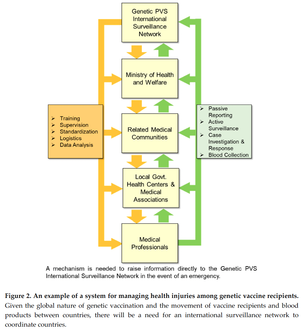
each country (Figure 2). It is expected that it will be important to incorporate not only the perspective
of infectious diseases but also biosafety and biosecurity [153,156].
As for Japan, Article 15 (2) of the Infectious Disease Act
(https://www.japaneselawtranslation.go.jp/ja/laws/view/2830/en#je_ch3at5) stipulates that the
Japanese government is responsible for conducting epidemiological studies. Given the significant
health risks associated with COVID-19 PVS, we urge the Japanese government to prioritize the
analysis and safety verification of blood products derived from gene vaccine recipients. This is
imperative given the urgent nature of the situation.
6. Other Important Considerations
There is an urgent need to develop methods to identify as well as remove spike proteins and
modified genes derived from gene vaccines in blood products. In order to develop a uniform
inspection standard, there is an urgent need in Japan for the Japanese Society of Hematology
(http://www.jshem.or.jp/modules/en/index.php?content_id=1), the Japanese Society of Transfusion
and Cell Therapy (http://yuketsu.jstmct.or.jp/en/), and their related organizations to develop
guidelines on how to handle blood products that contain residual spike proteins or their modified
genes. Also, as noted earlier, gene vaccination has been promoted on a global scale [2,3], which will
necessitate coordination and exchange of information with national administrations and relevant
international medical societies (Figure 1). International guidelines on the handling of blood products
and the establishment of an international investigatory organization will be necessary (Figure 2).
However, there is an urgent need to share the risks of transfusion of blood products derived from
genetic vaccine recipients among the parties concerned, and prompt investigation and response by
all parties concerned is essential. The most important initial action is to make the relevant medical
personnel aware of this situation.
In the development of various guidelines, it will be helpful to refer to the response of each
country when the transmission of BSE and vCJD, also through blood transfusion, became a problem
(e.g. the Creutzfeldt-Jakob Disease International Surveillance Network in
https://www.eurocjd.ed.ac.uk/) [110,114,115,121,157]. For example, in the United Kingdom, when
BSE became a social problem and the mode of transmission of prion protein was unknown,
leukodepletion of blood products was conducted universally. Whether this was effective in
preventing transmission of BSE and vCJD through blood products is controversial [110,120,121,158],
but it was not common at the time to remove white blood cells from all blood products, as is now
routinely done with collected blood. However, because of leukodepletion, the safety of blood
products has increased [159]. In the case of the spike protein, which causes abnormalities such as
agglutination of red blood cells and platelets [8–11,49], we do not expect the problem to be eliminated
by leukodepletion alone. However, it is worth confirming whether washing of red blood cells can be
effective [160,161]. In urgent cases, autotransfusion may be an option [162].
Recent studies have shown that RNA pseudouridylation can result in frameshifting [133]. It is
not yet clear whether a portion of the pseudouridinated mRNA for the spike protein is translated into
another protein of unknown function in vaccine recipients. If these proteins are also pathogenic,
additional testing for such frameshift proteins may be needed in the future. Even if a frameshift
protein is not toxic, it must be foreign to the body and could cause autoimmune disease. In addition,
LNPs themselves are highly inflammatory substances [23,100–102], as described in Section 3.1, but
LNPs have been found to have stronger adjuvant activity than the adjuvants used in conventional
vaccines [104], and there is also concern about autoimmune diseases resulting from this aspect (Table
1, point 4) [105,163]. Thus, although it is not clear what the causative agent of autoimmune disease
is, the large number of reported cases of autoimmune disease following genetic vaccination is
extremely concerning [15,21,27,30,105,164]. The very mechanism of gene vaccines that causes one’s
own cells to produce the antigens of pathogens carries the risk of inducing autoimmune diseases,
which cannot be completely avoided even if mRNA pseudouridylation technology is used. In this
context, individuals with a positive blood test for spike protein may need to have interviews and
additional tests for autoimmune disease indicators, such as antinuclear antibodies (Table 2, point 4)
[27,105,129,130]. Alternatively, if the amino acid sequence of the protein resulting from the frameshift
is predictable, these candidate proteins could be included in the initial mass spectrometry assay
(Table 2, point 6). In any case, it is particularly important to develop tests and establish medical care
settings in anticipation of these situations.
7. Conclusion
Finally, we would like to state that if we continue to use genetic vaccines such as
pseudouridinated mRNAs and mRNA-LNP platforms [46,103], there will be further risks like those
described in this review. It should also be stressed that the issues discussed here are matters that
pertain to all organ transplants, including bone marrow transplants, and not just blood products. The
impact of these genetic vaccines on blood products and the actual damage caused by them are
unknown at present. Therefore, in order to avoid these risks and prevent further expansion of blood
contamination and complication of the situation, we strongly request that the vaccination campaign
using genetic vaccines be suspended and that a harm–benefit assessment be carried out as early as
possible, as called for by Fraiman et al. and Polykretis et al. [27,31–33]. As we have repeatedly stated,
the health injuries caused by genetic vaccination are already extremely serious, and it is high time
that countries and relevant organizations take concrete steps together to identify the risks and to
control and resolve them.
Author Contributions: Conceptualization, J.U. M.F. and A.F.; investigation, J.U. H.M. Y.M. M.F. and A.F.;
resources, Y.H.; data curation, J.U. H.M. M.F. and A.F.; writing—original draft preparation, J.U.; writing—
review and editing, J.U. H.M. Y.H. K.Y. M.F. and A.F.; visualization, J.U.; supervision, J.U. M.F. and A.F.; project
administration, J.U. M.F. and A.F.; funding acquisition, M.F. and A.F. All authors have read and agreed to the
published version of the manuscript.
Funding: The study was supported by donations from members of the Japanese Society for Vaccine-related
Complications and the Volunteer Medical Association.
Institutional Review Board Statement: Not applicable.
Acknowledgments: We would like to express our deep appreciation to the members of the Volunteer Medical
Association for their help in the discussions that led to the preparation of this review.
Conflicts of Interest: The authors declare no conflict of interest in connection with this research.
References
1. Sohrabi, C.; Alsafi, Z.; O’Neill, N.; Khan, M.; Kerwan, A.; Al-Jabir, A.; Iosifidis, C.; Agha, R. World Health
Organization declares global emergency: A review of the 2019 novel coronavirus (COVID-19). International
Journal of Surgery 2020, 76, 71–76.
2. Francis, A.I.; Ghany, S.; Gilkes, T.; Umakanthan, S. Review of COVID-19 vaccine subtypes, efficacy and
geographical distributions. Postgraduate Medical Journal 2022, 98, 389–394.
3. Patel, R.; Kaki, M.; Potluri, V.S.; Kahar, P.; Khanna, D. A comprehensive review of SARS-CoV-2 vaccines:
Pfizer, Moderna & Johnson & Johnson. Human Vaccines & Immunotherapeutics 2022, 18.
4. Harrison, A.G.; Lin, T.; Wang, P. Mechanisms of SARS-CoV-2 Transmission and Pathogenesis. Trends in
Immunology 2020, 41, 1100–1115.
5. Lamers, M.M.; Haagmans, B.L. SARS-CoV-2 pathogenesis. Nature Reviews Microbiology 2022, 20, 270–284.
6. Lan, J.; Ge, J.; Yu, J.; Shan, S.; Zhou, H.; Fan, S.; Zhang, Q.; Shi, X.; Wang, Q.; Zhang, L.; Wang, X. Structure
of the SARS-CoV-2 spike receptor-binding domain bound to the ACE2 receptor. Nature 2020, 581, 215–220.
7. Zhang, S.; Liu, Y.; Wang, X.; Yang, L.; Li, H.; Wang, Y.; Liu, M.; Zhao, X.; Xie, Y.; Yang, Y.; Zhang, S.; Fan,
Z.; Dong, J.; Yuan, Z.; Ding, Z.; Zhang, Y.; Hu, L. SARS-CoV-2 binds platelet ACE2 to enhance thrombosis
in COVID-19. Journal of Hematology & Oncology 2020, 13.
8. Berzuini, A.; Bianco, C.; Migliorini, A.C.; Maggioni, M.; Valenti, L.; Prati, D. Red blood cell morphology in
patients with COVID-19-related anaemia. Blood Transfus 2021, 19, 34–36.
9. Melkumyants, A.; Buryachkovskaya, L.; Lomakin, N.; Antonova, O.; Serebruany, V. Mild COVID-19 and
Impaired Blood Cell–Endothelial Crosstalk: Considering Long-Term Use of Antithrombotics? Thrombosis
and Haemostasis 2021, 122, 123–130.
10. Boschi, C.; Scheim, D.E.; Bancod, A.; Militello, M.; Bideau, M.L.; Colson, P.; Fantini, J.; Scola, B.L. SARSCoV-
2 Spike Protein Induces Hemagglutination: Implications for COVID-19 Morbidities and Therapeutics
and for Vaccine Adverse Effects. International Journal of Molecular Sciences 2022, 23.
11. Scheim, D.E. A Deadly Embrace: Hemagglutination Mediated by SARS-CoV-2 Spike Protein at Its 22 NGlycosylation
Sites, Red Blood Cell Surface Sialoglycoproteins, and Antibody. International Journal of
Molecular Sciences 2022, 23.
12. McFadyen, J.D.; Stevens, H.; Peter, K. The Emerging Threat of (Micro)Thrombosis in COVID-19 and Its
Therapeutic Implications. Circulation Research 2020, 127, 571–587.
13. Grobbelaar, Lize M.; Venter, C.; Vlok, M.; Ngoepe, M.; Laubscher, Gert J.; Lourens, Petrus J.; Steenkamp,
J.; Kell, Douglas B.; Pretorius, E. SARS-CoV-2 spike protein S1 induces fibrin(ogen) resistant to fibrinolysis:
implications for microclot formation in COVID-19. Bioscience Reports 2021, 41.
14. Bilotta, C.; Perrone, G.; Adelfio, V.; Spatola, G.F.; Uzzo, M.L.; Argo, A.; Zerbo, S. COVID-19 Vaccine-Related
Thrombosis: A Systematic Review and Exploratory Analysis. Front Immunol 2021, 12, 729251.
15. Garg, R.K.; Paliwal, V.K. Spectrum of neurological complications following COVID-19 vaccination.
Neurological Sciences 2021, 43, 3–40.
16. Oldenburg, J.; Klamroth, R.; Langer, F.; Albisetti, M.; von Auer, C.; Ay, C.; Korte, W.; Scharf, R.E.; Pötzsch,
B.; Greinacher, A. Diagnosis and Management of Vaccine-Related Thrombosis following AstraZeneca
COVID-19 Vaccination: Guidance Statement from the GTH. Hämostaseologie 2021, 41, 184–189.
17. Sharifian-Dorche, M.; Bahmanyar, M.; Sharifian-Dorche, A.; Mohammadi, P.; Nomovi, M.; Mowla, A.
Vaccine-induced immune thrombotic thrombocytopenia and cerebral venous sinus thrombosis post
COVID-19 vaccination; a systematic review. J Neurol Sci 2021, 428, 117607.
18. Lane, S.; Yeomans, A.; Shakir, S. Reports of myocarditis and pericarditis following mRNA COVID-19
vaccination: a systematic review of spontaneously reported data from the UK, Europe and the USA and of
the scientific literature. BMJ Open 2022, 12.
19. Oster, M.E.; Shay, D.K.; Su, J.R.; Gee, J.; Creech, C.B.; Broder, K.R.; Edwards, K.; Soslow, J.H.; Dendy, J.M.;
Schlaudecker, E.; Lang, S.M.; Barnett, E.D.; Ruberg, F.L.; Smith, M.J.; Campbell, M.J.; Lopes, R.D.; Sperling,
L.S.; Baumblatt, J.A.; Thompson, D.L.; Marquez, P.L.; Strid, P.; Woo, J.; Pugsley, R.; Reagan-Steiner, S.;
DeStefano, F.; Shimabukuro, T.T. Myocarditis Cases Reported After mRNA-Based COVID-19 Vaccination
in the US From December 2020 to August 2021. Jama 2022, 327.
20. Yasmin, F.; Najeeb, H.; Naeem, U.; Moeed, A.; Atif, A.R.; Asghar, M.S.; Nimri, N.; Saleem, M.;
Bandyopadhyay, D.; Krittanawong, C.; Fadelallah Eljack, M.M.; Tahir, M.J.; Waqar, F. Adverse events
following COVID-19 mRNA vaccines: A systematic review of cardiovascular complication, thrombosis,
and thrombocytopenia. Immun Inflamm Dis 2023, 11, e807.
21. Konishi, N.; Hirai, Y.; Hikota, H.; Miyahara, S.; Fujisawa, A.; Motohashi, H.; Ueda, J.; Inoue, M.; Fukushima,
M. Quantifying side effects of COVID-19 vaccines: A PubMed survey of papers on diseases as side effects
presented at academic conferences in Japan. Rinsho Hyoka (Clinical Evaluation) 2024, 51.
22. Parry, P.I.; Lefringhausen, A.; Turni, C.; Neil, C.J.; Cosford, R.; Hudson, N.J.; Gillespie, J. ‘Spikeopathy’:
COVID-19 Spike Protein Is Pathogenic, from Both Virus and Vaccine mRNA. Biomedicines 2023, 11.
23. Ndeupen, S.; Qin, Z.; Jacobsen, S.; Bouteau, A.; Estanbouli, H.; Igyártó, B.Z. The mRNA-LNP platform’s
lipid nanoparticle component used in preclinical vaccine studies is highly inflammatory. iScience 2021, 24.
24. Maugeri, M.; Nawaz, M.; Papadimitriou, A.; Angerfors, A.; Camponeschi, A.; Na, M.; Hölttä, M.; Skantze,
P.; Johansson, S.; Sundqvist, M.; Lindquist, J.; Kjellman, T.; Mårtensson, I.-L.; Jin, T.; Sunnerhagen, P.;
Östman, S.; Lindfors, L.; Valadi, H. Linkage between endosomal escape of LNP-mRNA and loading into
EVs for transport to other cells. Nature Communications 2019, 10.
25. Bansal, S.; Perincheri, S.; Fleming, T.; Poulson, C.; Tiffany, B.; Bremner, R.M.; Mohanakumar, T. Cutting
Edge: Circulating Exosomes with COVID Spike Protein Are Induced by BNT162b2 (Pfizer–BioNTech)
Vaccination prior to Development of Antibodies: A Novel Mechanism for Immune Activation by mRNA
Vaccines. The Journal of Immunology 2021, 207, 2405–2410.
26. Seneff, S.; Nigh, G.; Kyriakopoulos, A.M.; McCullough, P.A. Innate immune suppression by SARS-CoV-2
mRNA vaccinations: The role of G-quadruplexes, exosomes, and MicroRNAs. Food Chem Toxicol 2022, 164,
113008.
27. Polykretis, P.; Donzelli, A.; Lindsay, J.C.; Wiseman, D.; Kyriakopoulos, A.M.; Mörz, M.; Bellavite, P.;
Fukushima, M.; Seneff, S.; McCullough, P.A. Autoimmune inflammatory reactions triggered by the
COVID-19 genetic vaccines in terminally differentiated tissues. Autoimmunity 2023, 56.
28. Brogna, C.; Cristoni, S.; Marino, G.; Montano, L.; Viduto, V.; Fabrowski, M.; Lettieri, G.; Piscopo, M.
Detection of recombinant Spike protein in the blood of individuals vaccinated against SARS-CoV-2:
Possible molecular mechanisms. Proteomics Clin Appl 2023, 17, e2300048.
29. Yonker, L.M.; Swank, Z.; Bartsch, Y.C.; Burns, M.D.; Kane, A.; Boribong, B.P.; Davis, J.P.; Loiselle, M.;
Novak, T.; Senussi, Y.; Cheng, C.A.; Burgess, E.; Edlow, A.G.; Chou, J.; Dionne, A.; Balaguru, D.; Lahoud-
Rahme, M.; Arditi, M.; Julg, B.; Randolph, A.G.; Alter, G.; Fasano, A.; Walt, D.R. Circulating Spike Protein
Detected in Post-COVID-19 mRNA Vaccine Myocarditis. Circulation 2023, 147, 867–876.
30. Chen, Y.; Xu, Z.; Wang, P.; Li, X.M.; Shuai, Z.W.; Ye, D.Q.; Pan, H.F. New-onset autoimmune phenomena
post-COVID-19 vaccination. Immunology 2022, 165, 386–401.
31. Polykretis, P.; McCullough, P.A. Rational harm-benefit assessments by age group are required for
continued COVID-19 vaccination. Scandinavian Journal of Immunology 2022, 98.
32. Fraiman, J.; Erviti, J.; Jones, M.; Greenland, S.; Whelan, P.; Kaplan, R.M.; Doshi, P. Serious adverse events
of special interest following mRNA COVID-19 vaccination in randomized trials in adults. Vaccine 2022, 40,
5798–5805.
33. Bardosh, K.; Krug, A.; Jamrozik, E.; Lemmens, T.; Keshavjee, S.; Prasad, V.; Makary, M.A.; Baral, S.; Høeg,
T.B. COVID-19 vaccine boosters for young adults: a risk benefit assessment and ethical analysis of mandate
policies at universities. Journal of Medical Ethics 2024, 50, 126–138.
34. Stanworth, S.J.; New, H.V.; Apelseth, T.O.; Brunskill, S.; Cardigan, R.; Doree, C.; Germain, M.; Goldman,
M.; Massey, E.; Prati, D.; Shehata, N.; So-Osman, C.; Thachil, J. Effects of the COVID-19 pandemic on supply
and use of blood for transfusion. The Lancet Haematology 2020, 7, e756–e64.
35. Chang, L.; Yan, Y.; Wang, L. Coronavirus Disease 2019: Coronaviruses and Blood Safety. Transfusion
Medicine Reviews 2020, 34, 75–80.
36. Bouhou, S.; Lahjouji, K.; Masrar, A. Blood donor eligibility after COVID-19 vaccination: the current state of
recommendations. Pan Afr Med J 2021, 40, 207.
37. Jacobs, J.W.; Bibb, L.A.; Savani, B.N.; Booth, G.S. Refusing blood transfusions from COVID-19-vaccinated
donors: are we repeating history? British Journal of Haematology 2021, 196, 585–588.
38. Hunain, R.; Uday, U.; Rackimuthu, S.; Nawaz, F.A.; Narain, K.; Essar, M.Y.; Rehman, M.U.; Ahmad, S.;
Butt, A. Effects of SARS-CoV-2 vaccination on blood donation and blood banks in India. Ann Med Surg
(Lond) 2022, 78, 103772.
39. Roubinian, N.H.; Greene, J.; Liu, V.X.; Lee, C.; Mark, D.G.; Vinson, D.R.; Spencer, B.R.; Bruhn, R.; Bravo,
M.; Stone, M.; Custer, B.; Kleinman, S.; Busch, M.P.; Norris, P.J. Clinical outcomes in hospitalized plasma
and platelet transfusion recipients prior to and following widespread blood donor SARS-CoV-2 infection
and vaccination. Transfusion 2023, 64, 53–67.
40. Fertig, T.E.; Chitoiu, L.; Marta, D.S.; Ionescu, V.-S.; Cismasiu, V.B.; Radu, E.; Angheluta, G.; Dobre, M.;
Serbanescu, A.; Hinescu, M.E.; Gherghiceanu, M. Vaccine mRNA Can Be Detected in Blood at 15 Days
Post-Vaccination. Biomedicines 2022, 10.
41. Mörz, M. A Case Report: Multifocal Necrotizing Encephalitis and Myocarditis after BNT162b2 mRNA
Vaccination against COVID-19. Vaccines 2022, 10.
42. Yamamoto, M.; Kase, M.; Sano, H.; Kamijima, R.; Sano, S. Persistent varicella zoster virus infection
following mRNA COVID-19 vaccination was associated with the presence of encoded spike protein in the
lesion. Journal of Cutaneous Immunology and Allergy 2022, 6, 18–23.
43. Castruita, J.A.S.; Schneider, U.V.; Mollerup, S.; Leineweber, T.D.; Weis, N.; Bukh, J.; Pedersen, M.S.; Westh,
H. SARS-CoV-2 spike mRNA vaccine sequences circulate in blood up to 28 days after COVID-19
vaccination. APMIS 2023, 131, 128–132.
44. Krauson, A.J.; Casimero, F.V.C.; Siddiquee, Z.; Stone, J.R. Duration of SARS-CoV-2 mRNA vaccine
persistence and factors associated with cardiac involvement in recently vaccinated patients. NPJ Vaccines
2023, 8, 141.
45. Xu, S.; Yang, K.; Li, R.; Zhang, L. mRNA Vaccine Era-Mechanisms, Drug Platform and Clinical Prospection.
Int J Mol Sci 2020, 21.
46. Bitounis, D.; Jacquinet, E.; Rogers, M.A.; Amiji, M.M. Strategies to reduce the risks of mRNA drug and
vaccine toxicity. Nat Rev Drug Discov 2024.
47. Yamamoto, K. Adverse effects of COVID-19 vaccines and measures to prevent them. Virology Journal 2022,
19.
48. Rodriguez, Y.; Rojas, M.; Beltran, S.; Polo, F.; Camacho-Dominguez, L.; Morales, S.D.; Gershwin, M.E.;
Anaya, J.M. Autoimmune and autoinflammatory conditions after COVID-19 vaccination. New case reports
and updated literature review. J Autoimmun 2022, 132, 102898.
49. Perico, L.; Morigi, M.; Galbusera, M.; Pezzotta, A.; Gastoldi, S.; Imberti, B.; Perna, A.; Ruggenenti, P.;
Donadelli, R.; Benigni, A.; Remuzzi, G. SARS-CoV-2 Spike Protein 1 Activates Microvascular Endothelial
Cells and Complement System Leading to Platelet Aggregation. Front Immunol 2022, 13, 827146.
50. Idrees, D.; Kumar, V. SARS-CoV-2 spike protein interactions with amyloidogenic proteins: Potential clues
to neurodegeneration. Biochemical and Biophysical Research Communications 2021, 554, 94–98.
51. Charnley, M.; Islam, S.; Bindra, G.K.; Engwirda, J.; Ratcliffe, J.; Zhou, J.; Mezzenga, R.; Hulett, M.D.; Han,
K.; Berryman, J.T.; Reynolds, N.P. Neurotoxic amyloidogenic peptides in the proteome of SARS-COV2:
potential implications for neurological symptoms in COVID-19. Nature Communications 2022, 13.
52. Kruger, A.; Vlok, M.; Turner, S.; Venter, C.; Laubscher, G.J.; Kell, D.B.; Pretorius, E. Proteomics of fibrin
amyloid microclots in long COVID/post-acute sequelae of COVID-19 (PASC) shows many entrapped proinflammatory
molecules that may also contribute to a failed fibrinolytic system. Cardiovascular Diabetology
2022, 21.
53. Nyström, S.; Hammarström, P. Amyloidogenesis of SARS-CoV-2 Spike Protein. Journal of the American
Chemical Society 2022, 144, 8945–8950.
54. Chesney, A.D.; Maiti, B.; Hansmann, U.H.E. SARS-COV-2 spike protein fragment eases amyloidogenesis
of alpha-synuclein. J Chem Phys 2023, 159.
55. Olajide, O.A.; Iwuanyanwu, V.U.; Adegbola, O.D.; Al-Hindawi, A.A. SARS-CoV-2 Spike Glycoprotein S1
Induces Neuroinflammation in BV-2 Microglia. Molecular Neurobiology 2021, 59, 445–458.
56. Oh, J.; Cho, W.-H.; Barcelon, E.; Kim, K.H.; Hong, J.; Lee, S.J. SARS-CoV-2 spike protein induces cognitive
deficit and anxiety-like behavior in mouse via non-cell autonomous hippocampal neuronal death. Scientific
Reports 2022, 12.
57. O’Brien, B.C.V.; Weber, L.; Hueffer, K.; Weltzin, M.M. SARS-CoV-2 spike ectodomain targets alpha7
nicotinic acetylcholine receptors. J Biol Chem 2023, 299, 104707.
58. Buzhdygan, T.P.; DeOre, B.J.; Baldwin-Leclair, A.; Bullock, T.A.; McGary, H.M.; Khan, J.A.; Razmpour, R.;
Hale, J.F.; Galie, P.A.; Potula, R.; Andrews, A.M.; Ramirez, S.H. The SARS-CoV-2 spike protein alters barrier
function in 2D static and 3D microfluidic in-vitro models of the human blood-brain barrier. Neurobiol Dis
2020, 146, 105131.
59. Rhea, E.M.; Logsdon, A.F.; Hansen, K.M.; Williams, L.M.; Reed, M.J.; Baumann, K.K.; Holden, S.J.; Raber,
J.; Banks, W.A.; Erickson, M.A. The S1 protein of SARS-CoV-2 crosses the blood–brain barrier in mice.
Nature Neuroscience 2020, 24, 368–378.
60. Zhang, L.; Zhou, L.; Bao, L.; Liu, J.; Zhu, H.; Lv, Q.; Liu, R.; Chen, W.; Tong, W.; Wei, Q.; Xu, Y.; Deng, W.;
Gao, H.; Xue, J.; Song, Z.; Yu, P.; Han, Y.; Zhang, Y.; Sun, X.; Yu, X.; Qin, C. SARS-CoV-2 crosses the blood–
brain barrier accompanied with basement membrane disruption without tight junctions alteration. Signal
Transduction and Targeted Therapy 2021, 6.
61. Trougakos, I.P.; Terpos, E.; Alexopoulos, H.; Politou, M.; Paraskevis, D.; Scorilas, A.; Kastritis, E.;
Andreakos, E.; Dimopoulos, M.A. Adverse effects of COVID-19 mRNA vaccines: the spike hypothesis.
Trends in Molecular Medicine 2022, 28, 542–554.
62. Halma, M.T.J.; Plothe, C.; Marik, P.; Lawrie, T.A. Strategies for the Management of Spike Protein-Related
Pathology. Microorganisms 2023, 11.
63. Monge, S.; Pastor-Barriuso, R.; Hernán, M.A. The imprinting effect of covid-19 vaccines: an expected
selection bias in observational studies. Bmj 2023.
64. Wang, Q.; Guo, Y.; Tam, A.R.; Valdez, R.; Gordon, A.; Liu, L.; Ho, D.D. Deep immunological imprinting
due to the ancestral spike in the current bivalent COVID-19 vaccine. Cell Rep Med 2023, 4, 101258.
65. Shrestha, N.K.; Burke, P.C.; Nowacki, A.S.; Simon, J.F.; Hagen, A.; Gordon, S.M. Effectiveness of the
Coronavirus Disease 2019 Bivalent Vaccine. Open Forum Infectious Diseases 2023, 10.
66. Arvin, A.M.; Fink, K.; Schmid, M.A.; Cathcart, A.; Spreafico, R.; Havenar-Daughton, C.; Lanzavecchia, A.;
Corti, D.; Virgin, H.W. A perspective on potential antibody-dependent enhancement of SARS-CoV-2.
Nature 2020, 584, 353–363.
67. Lee, W.S.; Wheatley, A.K.; Kent, S.J.; DeKosky, B.J. Antibody-dependent enhancement and SARS-CoV-2
vaccines and therapies. Nat Microbiol 2020, 5, 1185–1191.
68. Irrgang, P.; Gerling, J.; Kocher, K.; Lapuente, D.; Steininger, P.; Habenicht, K.; Wytopil, M.; Beileke, S.;
Schäfer, S.; Zhong, J.; Ssebyatika, G.; Krey, T.; Falcone, V.; Schülein, C.; Peter, A.S.; Nganou-Makamdop,
K.; Hengel, H.; Held, J.; Bogdan, C.; Überla, K.; Schober, K.; Winkler, T.H.; Tenbusch, M. Class switch
toward noninflammatory, spike-specific IgG4 antibodies after repeated SARS-CoV-2 mRNA vaccination.
Science Immunology 2023, 8.
69. Kiszel, P.; Sík, P.; Miklós, J.; Kajdácsi, E.; Sinkovits, G.; Cervenak, L.; Prohászka, Z. Class switch towards
spike protein-specific IgG4 antibodies after SARS-CoV-2 mRNA vaccination depends on prior infection
history. Scientific Reports 2023, 13.
70. Uversky, V.; Redwan, E.; Makis, W.; Rubio-Casillas, A. IgG4 Antibodies Induced by Repeated Vaccination
May Generate Immune Tolerance to the SARS-CoV-2 Spike Protein. Vaccines 2023, 11.
71. Yoshimura, M.; Sakamoto, A.; Ozuru, R.; Kurihara, Y.; Itoh, R.; Ishii, K.; Shimizu, A.; Chou, B.; Nabeshima,
S.; Hiromatsu, K. The appearance of anti-spike receptor binding domain immunoglobulin G4 responses
after repetitive immunization with messenger RNA-based COVID-19 vaccines. Int J Infect Dis 2024, 139, 1–
5.
72. Murata, K.; Nakao, N.; Ishiuchi, N.; Fukui, T.; Katsuya, N.; Fukumoto, W.; Oka, H.; Yoshikawa, N.; Nagao,
T.; Namera, A.; Kakimoto, N.; Oue, N.; Awai, K.; Yoshimoto, K.; Nagao, M. Four cases of cytokine storm
after COVID-19 vaccination: Case report. Front Immunol 2022, 13, 967226.
73. Masset, C.; Kervella, D.; Kandel-Aznar, C.; Fantou, A.; Blancho, G.; Hamidou, M. Relapse of IgG4-related
nephritis following mRNA COVID-19 vaccine. Kidney International 2021, 100, 465–466.
74. Patel, A.H. Acute Liver Injury and IgG4-related Autoimmune Pancreatitis following mRNA based COVID-
19 vaccination. Hepatology Forum 2022.
75. Aochi, S.; Uehara, M.; Yamamoto, M. IgG4-related Disease Emerging after COVID-19 mRNA Vaccination.
Internal Medicine 2023, 62, 1547–1551.
76. Katsikas Triantafyllidis, K.; Giannos, P.; Mian, I.T.; Kyrtsonis, G.; Kechagias, K.S. Varicella Zoster Virus
Reactivation Following COVID-19 Vaccination: A Systematic Review of Case Reports. Vaccines 2021, 9.
77. Lensen, R.; Netea, M.G.; Rosendaal, F.R. Hepatitis C Virus Reactivation Following COVID-19
Vaccination—A Case Report. Int Med Case Rep J 2021, 14, 573–576.
78. Psichogiou, M.; Samarkos, M.; Mikos, N.; Hatzakis, A. Reactivation of Varicella Zoster Virus after
Vaccination for SARS-CoV-2. Vaccines 2021, 9.
79. Fathy, R.A.; McMahon, D.E.; Lee, C.; Chamberlin, G.C.; Rosenbach, M.; Lipoff, J.B.; Tyagi, A.; Desai, S.R.;
French, L.E.; Lim, H.W.; Thiers, B.H.; Hruza, G.J.; Fassett, M.; Fox, L.P.; Greenberg, H.L.; Blumenthal, K.;
Freeman, E.E. Varicella-zoster and herpes simplex virus reactivation post-COVID-19 vaccination: a review
of 40 cases in an International Dermatology Registry. J Eur Acad Dermatol Venereol 2022, 36, e6–e9.
80. Gringeri, M.; Battini, V.; Cammarata, G.; Mosini, G.; Guarnieri, G.; Leoni, C.; Pozzi, M.; Radice, S.; Clementi,
E.; Carnovale, C. Herpes zoster and simplex reactivation following COVID-19 vaccination: new insights
from a vaccine adverse event reporting system (VAERS) database analysis. Expert Rev Vaccines 2022, 21,
675–684.
81. Hertel, M.; Heiland, M.; Nahles, S.; von Laffert, M.; Mura, C.; Bourne, P.E.; Preissner, R.; Preissner, S. Realworld
evidence from over one million COVID-19 vaccinations is consistent with reactivation of the
varicella-zoster virus. Journal of the European Academy of Dermatology and Venereology 2022, 36, 1342–1348.
82. Shafiee, A.; Amini, M.J.; Arabzadeh Bahri, R.; Jafarabady, K.; Salehi, S.A.; Hajishah, H.; Mozhgani, S.-H.
Herpesviruses reactivation following COVID-19 vaccination: a systematic review and meta-analysis.
European Journal of Medical Research 2023, 28.
83. Culver, J. Preventing transmission of blood-borne pathogens: a compelling argument for effective deviceselection
strategies. Am J Infect Control 1997, 25, 430–433.
84. Beltrami, E.M.; Williams, I.T.; Shapiro, C.N.; Chamberland, M.E. Risk and Management of Blood-Borne
Infections in Health Care Workers. Clinical Microbiology Reviews 2000, 13, 385–407.
85. Ison, M.G.; Grossi, P.; Practice, A.S.T.I.D.C. o. Donor-derived infections in solid organ transplantation. Am
J Transplant 2013, 13 Suppl 4, 22-30.
86. Fishman, J.A.; Grossi, P.A. Donor-derived infection--the challenge for transplant safety. Nat Rev Nephrol
2014, 10, 663–672.
87. Bahakel, H.K.; Pellet Madan, R.; Danziger-Isakov, L. Approach to suspected donor-derived infections.
Front Pediatr 2023, 11, 1265023.
88. Tobin, G.J.; Trujillo, J.D.; Bushnell, R.V.; Lin, G.; Chaudhuri, A.R.; Long, J.; Barrera, J.; Pena, L.; Grubman,
M.J.; Nara, P.L. Deceptive imprinting and immune refocusing in vaccine design. Vaccine 2008, 26, 6189–
6199.
89. Gatto, D.; Brink, R. The germinal center reaction. J Allergy Clin Immunol 2010, 126, 898–907; quiz 08-9.
90. Seneff, S.; Nigh, G. Worse Than the Disease? Reviewing Some Possible Unintended Consequences of the
mRNA Vaccines Against COVID-19. International Journal of Vaccine Theory, Practice, and Research 2021, 2, 38–
79.
91. Bernardini, A.; Gigli, G.L.; Janes, F.; Pellitteri, G.; Ciardi, C.; Fabris, M.; Valente, M. Creutzfeldt-Jakob
disease after COVID-19: infection-induced prion protein misfolding? A case report. Prion 2022, 16, 78–83.
92. Lukiw, W.J.; Jaber, V.R.; Pogue, A.I.; Zhao, Y. SARS-CoV-2 Invasion and Pathological Links to Prion
Disease. Biomolecules 2022, 12.
93. Tetz, G.; Tetz, V. Prion-like Domains in Spike Protein of SARS-CoV-2 Differ across Its Variants and Enable
Changes in Affinity to ACE2. Microorganisms 2022, 10.
94. Makhoul, K.; Beeber, T.; Cordero, R.; Khan, A.; Saliaj, M. Prion Disease After COVID-19: A Case Report.
Am J Case Rep 2023, 24, e940564.
95. Perez, J.-C.; Moret-Chalmin, C.; Montagnier, L. Emergence of a New Creutzfeldt-Jakob Disease: 26 Cases
of the Human Version of Mad-Cow Disease, Days After a COVID-19 Injection. International Journal of
Vaccine Theory, Practice, and Research 2023, 3, 727–770.
96. Seneff, S.; Kyriakopoulos, A.M.; Nigh, G.; McCullough, P.A. A Potential Role of the Spike Protein in
Neurodegenerative Diseases: A Narrative Review. Cureus 2023.
97. Perez, J.C.; Lounnas, V.; Montagnier, M. The Omicron Variant Breaks the Evolutionary Lineage of Sars-
Cov2 Variants. International Journal of Research -GRANTHAALAYAH 2021, 9, 108–132.
98. Bhardwaj, T.; Gadhave, K.; Kapuganti, S.K.; Kumar, P.; Brotzakis, Z.F.; Saumya, K.U.; Nayak, N.; Kumar,
A.; Joshi, R.; Mukherjee, B.; Bhardwaj, A.; Thakur, K.G.; Garg, N.; Vendruscolo, M.; Giri, R. Amyloidogenic
proteins in the SARS-CoV and SARS-CoV-2 proteomes. Nature Communications 2023, 14.
99. Faizullin, D.; Valiullina, Y.; Salnikov, V.; Zuev, Y. Direct interaction of fibrinogen with lipid microparticles
modulates clotting kinetics and clot structure. Nanomedicine 2020, 23, 102098.
100. Moghimi, S.M. Allergic Reactions and Anaphylaxis to LNP-Based COVID-19 Vaccines. Molecular Therapy
2021, 29, 898–900.
101. Moghimi, S.M.; Simberg, D. Pro-inflammatory concerns with lipid nanoparticles. Molecular Therapy 2022,
30, 2109–2110.
102. Tahtinen, S.; Tong, A.-J.; Himmels, P.; Oh, J.; Paler-Martinez, A.; Kim, L.; Wichner, S.; Oei, Y.; McCarron,
M.J.; Freund, E.C.; Amir, Z.A.; de la Cruz, C.C.; Haley, B.; Blanchette, C.; Schartner, J.M.; Ye, W.; Yadav,
M.; Sahin, U.; Delamarre, L.; Mellman, I. IL-1 and IL-1ra are key regulators of the inflammatory response
to RNA vaccines. Nature Immunology 2022, 23, 532–542.
103. Halma, M.T.J.; Rose, J.; Lawrie, T. The Novelty of mRNA Viral Vaccines and Potential Harms: A Scoping
Review. J 2023, 6, 220–235.
104. Alameh, M.G.; Tombacz, I.; Bettini, E.; Lederer, K.; Sittplangkoon, C.; Wilmore, J.R.; Gaudette, B.T.;
Soliman, O.Y.; Pine, M.; Hicks, P.; Manzoni, T.B.; Knox, J.J.; Johnson, J.L.; Laczko, D.; Muramatsu, H.; Davis,
B.; Meng, W.; Rosenfeld, A.M.; Strohmeier, S.; Lin, P.J.C.; Mui, B.L.; Tam, Y.K.; Kariko, K.; Jacquet, A.;
Krammer, F.; Bates, P.; Cancro, M.P.; Weissman, D.; Luning Prak, E.T.; Allman, D.; Locci, M.; Pardi, N.
Lipid nanoparticles enhance the efficacy of mRNA and protein subunit vaccines by inducing robust T
follicular helper cell and humoral responses. Immunity 2021, 54, 2877–2892 e7.
105. Jara, L.J.; Vera-Lastra, O.; Mahroum, N.; Pineda, C.; Shoenfeld, Y. Autoimmune post-COVID vaccine
syndromes: does the spectrum of autoimmune/inflammatory syndrome expand? Clinical Rheumatology
2022, 41, 1603–1609.
106. Varghese, J.L.; Fung, A.W.S.; Mattman, A.; Quach, T.T.T.; Gauiran, D.T.V.; Carruthers, M.N.; Chen, L.Y.C.
Clinical utility of serum IgG4 measurement. Clin Chim Acta 2020, 506, 228–235.
107. Katz, G.; Stone, J.H. Clinical Perspectives on IgG4-Related Disease and Its Classification. Annu Rev Med
2022, 73, 545–562.
108. Chapman, C.W. S. Project risk management: Process, techniques and insight. Wiley: London, UK, 2003.
109. Aven, T. Risk assessment and risk management: Review of recent advances on their foundation. European
Journal of Operational Research 2016, 253, 1–13.
110. Watson, N.; Brandel, J.-P.; Green, A.; Hermann, P.; Ladogana, A.; Lindsay, T.; Mackenzie, J.; Pocchiari, M.;
Smith, C.; Zerr, I.; Pal, S. The importance of ongoing international surveillance for Creutzfeldt–Jakob
disease. Nature Reviews Neurology 2021, 17, 362–379.
111. Maltezou, H.C.; Pavli, A.; Tsakris, A. Post-COVID Syndrome: An Insight on Its Pathogenesis. Vaccines 2021,
9.
112. Theoharides, T.C. Could SARS-CoV-2 Spike Protein Be Responsible for Long-COVID Syndrome? Molecular
Neurobiology 2022, 59, 1850–1861.
113. Greene, C.; Connolly, R.; Brennan, D.; Laffan, A.; O’Keeffe, E.; Zaporojan, L.; O’Callaghan, J.; Thomson, B.;
Connolly, E.; Argue, R.; Martin-Loeches, I.; Long, A.; Cheallaigh, C.N.; Conlon, N.; Doherty, C.P.;
Campbell, M. Blood-brain barrier disruption and sustained systemic inflammation in individuals with long
COVID-associated cognitive impairment. Nat Neurosci 2024.
114. Houston, F.; Foster, J.D.; Chong, A.; Hunter, N.; Bostock, C.J. Transmission of BSE by blood transfusion in
sheep. Lancet 2000, 356, 999–1000.
115. Hunter, N.; Foster, J.; Chong, A.; McCutcheon, S.; Parnham, D.; Eaton, S.; MacKenzie, C.; Houston, F.
Transmission of prion diseases by blood transfusion. J Gen Virol 2002, 83, (Pt 11), 2897–2905.
116. Seki, Y.; Yamazaki, Y.; Inoue, Y.; Wakabayashi, C.; Seto, S. How HIV infected haemophiliacs in Japan were
informed of their HIV-positive status. AIDS Care 2002, 14, 651–664.
117. Llewelyn, C.A.; Hewitt, P.E.; Knight, R.S.; Amar, K.; Cousens, S.; Mackenzie, J.; Will, R.G. Possible
transmission of variant Creutzfeldt-Jakob disease by blood transfusion. Lancet 2004, 363, 417–421.
118. Cullinane, J. Tainted Blood and Vengeful Spirits: The Legacy of Japan’s Yakugai Eizu (AIDS) Trial. Culture,
Medicine and Psychiatry 2005, 29, 5–31.
119. Hewitt, P.E.; Llewelyn, C.A.; Mackenzie, J.; Will, R.G. Creutzfeldt–Jakob disease and blood transfusion:
results of the UK Transfusion Medicine Epidemiological Review study. Vox Sanguinis 2006, 91, 221–230.
120. McLeod, N.P.; Nugent, P.; Dixon, D.; Dennis, M.; Cornwall, M.; Mallinson, G.; Watkins, N.; Thomas, S.;
Sutton, J.M. Evaluation of efficacy of prion reduction filters using blood from an endogenously infected
263K scrapie hamster model. Transfusion 2015, 55, 2390–2397.
121. Seed, C.R.; Hewitt, P.E.; Dodd, R.Y.; Houston, F.; Cervenakova, L. Creutzfeldt-Jakob disease and blood
transfusion safety. Vox Sanguinis 2018, 113, 220–231.
122. Tighe, P.J.; Ryder, R.R.; Todd, I.; Fairclough, L.C. ELISA in the multiplex era: Potentials and pitfalls.
PROTEOMICS—Clinical Applications 2015, 9, (3–4), 406–422.
123. Macklin, A.; Khan, S.; Kislinger, T. Recent advances in mass spectrometry based clinical proteomics:
applications to cancer research. Clinical Proteomics 2020, 17.
124. Zhou, B.; Xu, K.; Zheng, X.; Chen, T.; Wang, J.; Song, Y.; Shao, Y.; Zheng, S. Application of exosomes as
liquid biopsy in clinical diagnosis. Signal Transduction and Targeted Therapy 2020, 5.
125. Wang, D.; Baudys, J.; Bundy, J.L.; Solano, M.; Keppel, T.; Barr, J.R. Comprehensive Analysis of the Glycan
Complement of SARS-CoV-2 Spike Proteins Using Signature Ions-Triggered Electron-Transfer/Higher-
Energy Collisional Dissociation (EThcD) Mass Spectrometry. Analytical Chemistry 2020, 92, 14730–14739.
126. Ding, Z.; Wang, N.; Ji, N.; Chen, Z.-S. Proteomics technologies for cancer liquid biopsies. Molecular Cancer
2022, 21.
127. Pu, R.; Liu, S.; Ren, X.; Shi, D.; Ba, Y.; Huo, Y.; Zhang, W.; Ma, L.; Liu, Y.; Yang, Y.; Cheng, N. The screening
value of RT-LAMP and RT-PCR in the diagnosis of COVID-19: systematic review and meta-analysis. J Virol
Methods 2022, 300, 114392.
128. Mustafa Hellou, M.; Górska, A.; Mazzaferri, F.; Cremonini, E.; Gentilotti, E.; De Nardo, P.; Poran, I.;
Leeflang, M.M.; Tacconelli, E.; Paul, M. Nucleic acid amplification tests on respiratory samples for the
diagnosis of coronavirus infections: a systematic review and meta-analysis. Clinical Microbiology and
Infection 2021, 27, 341–351.
129. Agmon-Levin, N.; Damoiseaux, J.; Kallenberg, C.; Sack, U.; Witte, T.; Herold, M.; Bossuyt, X.; Musset, L.;
Cervera, R.; Plaza-Lopez, A.; Dias, C.; Sousa, M.J.; Radice, A.; Eriksson, C.; Hultgren, O.; Viander, M.;
Khamashta, M.; Regenass, S.; Andrade, L.E.C.; Wiik, A.; Tincani, A.; Rönnelid, J.; Bloch, D.B.; Fritzler, M.J.;
Chan, E.K.L.; Garcia-De La Torre, I.; Konstantinov, K.N.; Lahita, R.; Wilson, M.; Vainio, O.; Fabien, N.;
Sinico, R.A.; Meroni, P.; Shoenfeld, Y. International recommendations for the assessment of autoantibodies
to cellular antigens referred to as anti-nuclear antibodies. Annals of the Rheumatic Diseases 2014, 73, 17–23.
130. Xiao, Z.X.; Miller, J.S.; Zheng, S.G. An updated advance of autoantibodies in autoimmune diseases.
Autoimmun Rev 2021, 20, 102743.
131. Tsang, S.; Royse, C.F.; Terkawi, A.S. Guidelines for developing, translating, and validating a questionnaire
in perioperative and pain medicine. Saudi J Anaesth 2017, 11, (Suppl 1), S80-S89.
132. Semmler, A.; Mundorf, A.K.; Kuechler, A.S.; Schulze-Bosse, K.; Heidecke, H.; Schulze-Forster, K.; Schott,
M.; Uhrberg, M.; Weinhold, S.; Lackner, K.J.; Pawlitzki, M.; Meuth, S.G.; Boege, F.; Ruhrländer, J. Chronic
Fatigue and Dysautonomia following COVID-19 Vaccination Is Distinguished from Normal Vaccination
Response by Altered Blood Markers. Vaccines 2023, 11.
133. Mulroney, T.E.; Pöyry, T.; Yam-Puc, J.C.; Rust, M.; Harvey, R.F.; Kalmar, L.; Horner, E.; Booth, L.; Ferreira,
A.P.; Stoneley, M.; Sawarkar, R.; Mentzer, A.J.; Lilley, K.S.; Smales, C.M.; von der Haar, T.; Turtle, L.;
Dunachie, S.; Klenerman, P.; Thaventhiran, J.E.D.; Willis, A.E. N1-methylpseudouridylation of mRNA
causes +1 ribosomal frameshifting. Nature 2023.
134. Islam, A.; Bashir, M.S.; Joyce, K.; Rashid, H.; Laher, I.; Elshazly, S. An Update on COVID-19 Vaccine
Induced Thrombotic Thrombocytopenia Syndrome and Some Management Recommendations. Molecules
2021, 26.
135. Schaffner, A.; Risch, L.; Weber, M.; Thiel, S.; Jungert, K.; Pichler, M.; Wohlwend, N.; Lung, T.; Ritzler, M.;
Hillmann, D.; Copeland, S.; Renz, H.; Paprotny, M.; Risch, M. Sustained SARS-CoV-2 nucleocapsid
antibody levels in nonsevere COVID-19: a population-based study. Clin Chem Lab Med 2020, 59, e49–e51.
136. Chansaenroj, J.; Yorsaeng, R.; Posuwan, N.; Puenpa, J.; Wanlapakorn, N.; Sudhinaraset, N.; Sripramote, M.;
Chalongviriyalert, P.; Jirajariyavej, S.; Kiatpanabhikul, P.; Saiyarin, J.; Soudon, C.; Thienfaidee, O.;
Palakawong Na Ayuthaya, T.; Brukesawan, C.; Chirathaworn, C.; Intharasongkroh, D.; Chaiwanichsiri, D.;
Issarasongkhram, M.; Kitphati, R.; Mungaomklang, A.; Nagavajara, P.; Poovorawan, Y. Long-term specific
IgG response to SARS-CoV-2 nucleocapsid protein in recovered COVID-19 patients. Sci Rep 2021, 11, 23216.
137. Van Elslande, J.; Oyaert, M.; Ailliet, S.; Van Ranst, M.; Lorent, N.; Vande Weygaerde, Y.; Andre, E.; Lagrou,
K.; Vandendriessche, S.; Vermeersch, P. Longitudinal follow-up of IgG anti-nucleocapsid antibodies in
SARS-CoV-2 infected patients up to eight months after infection. J Clin Virol 2021, 136, 104765.
138. Mevorach, D.; Anis, E.; Cedar, N.; Bromberg, M.; Haas, E.J.; Nadir, E.; Olsha-Castell, S.; Arad, D.; Hasin,
T.; Levi, N.; Asleh, R.; Amir, O.; Meir, K.; Cohen, D.; Dichtiar, R.; Novick, D.; Hershkovitz, Y.; Dagan, R.;
Leitersdorf, I.; Ben-Ami, R.; Miskin, I.; Saliba, W.; Muhsen, K.; Levi, Y.; Green, M.S.; Keinan-Boker, L.;
Alroy-Preis, S. Myocarditis after BNT162b2 mRNA Vaccine against Covid-19 in Israel. New England Journal
of Medicine 2021, 385, 2140–2149.
139. Nakahara, T.; Iwabuchi, Y.; Miyazawa, R.; Tonda, K.; Shiga, T.; Strauss, H.W.; Antoniades, C.; Narula, J.;
Jinzaki, M. Assessment of Myocardial (18)F-FDG Uptake at PET/CT in Asymptomatic SARS-CoV-2-
vaccinated and Nonvaccinated Patients. Radiology 2023, 308, e230743.
140. Faksova, K.; Walsh, D.; Jiang, Y.; Griffin, J.; Phillips, A.; Gentile, A.; Kwong, J.C.; Macartney, K.; Naus, M.;
Grange, Z.; Escolano, S.; Sepulveda, G.; Shetty, A.; Pillsbury, A.; Sullivan, C.; Naveed, Z.; Janjua, N.Z.;
Giglio, N.; Perala, J.; Nasreen, S.; Gidding, H.; Hovi, P.; Vo, T.; Cui, F.; Deng, L.; Cullen, L.; Artama, M.;
Weintraub, E.; Lu, H.; Clothier, H.J.; Batty, K.; Paynter, J.; Petousis-Harris, H.; Buttery, J.; Black, S.; Hviid,
A. COVID-19 vaccines and adverse events of special interest: A multinational Global Vaccine Data Network
(GVDN) cohort study of 99 million vaccinated individuals. Vaccine 2024.
141. Pan, K.M.; Baldwin, M.; Nguyen, J.; Gasset, M.; Serban, A.; Groth, D.; Mehlhorn, I.; Huang, Z.; Fletterick,
R.J.; Cohen, F.E. Conversion of alpha-helices into beta-sheets features in the formation of the scrapie prion
proteins. Proceedings of the National Academy of Sciences 1993, 90, 10962–10966.
142. Langeveld, Jan P. M.; Wang, J.J.; Van de Wiel, Dick F. M.; Shih, Giles C.; Garssen, G.J.; Bossers, A.; Shih,
Jason C. H. Enzymatic Degradation of Prion Protein in Brain Stem from Infected Cattle and Sheep. The
Journal of Infectious Diseases 2003, 188, 1782–1789.
143. Prusiner, S.B.; Groth, D.F.; McKinley, M.P.; Cochran, S.P.; Bowman, K.A.; Kasper, K.C. Thiocyanate and
hydroxyl ions inactivate the scrapie agent. Proceedings of the National Academy of Sciences 1981, 78, 4606–
4610.
144. Race, R.E.; Raymond, G.J. Inactivation of Transmissible Spongiform Encephalopathy (Prion) Agents by
Environ LpH. Journal of Virology 2004, 78, 2164–2165.
145. Peretz, D.; Supattapone, S.; Giles, K.; Vergara, J.; Freyman, Y.; Lessard, P.; Safar, J.G.; Glidden, D.V.;
McCulloch, C.; Nguyen, H.-O. B.; Scott, M.; DeArmond, S.J.; Prusiner, S.B. Inactivation of Prions by Acidic
Sodium Dodecyl Sulfate. Journal of Virology 2006, 80, 322–331.
146. Stroup, D.F.; Berlin, J.A.; Morton, S.C.; Olkin, I.; Williamson, G.D.; Rennie, D.; Moher, D.; Becker, B.J.; Sipe,
T.A.; Thacker, S.B. Meta-analysis of observational studies in epidemiology: a proposal for reporting. Metaanalysis
Of Observational Studies in Epidemiology (MOOSE) group. JAMA 2000, 283, 2008–2012.
147. Moher, D.; Liberati, A.; Tetzlaff, J.; Altman, D.G.; Group, P. Preferred reporting items for systematic reviews
and meta-analyses: the PRISMA statement. Int J Surg 2010, 8, 336–341.
148. Murad, M.H.; Montori, V.M.; Ioannidis, J.P.; Jaeschke, R.; Devereaux, P.J.; Prasad, K.; Neumann, I.;
Carrasco-Labra, A.; Agoritsas, T.; Hatala, R.; Meade, M.O.; Wyer, P.; Cook, D.J.; Guyatt, G. How to read a
systematic review and meta-analysis and apply the results to patient care: users’ guides to the medical
literature. JAMA 2014, 312, 171–179.
149. Gould, C.V.; Free, R.J.; Bhatnagar, J.; Soto, R.A.; Royer, T.L.; Maley, W.R.; Moss, S.; Berk, M.A.; Craig-
Shapiro, R.; Kodiyanplakkal, R.P.L.; Westblade, L.F.; Muthukumar, T.; Puius, Y.A.; Raina, A.; Hadi, A.;
Gyure, K.A.; Trief, D.; Pereira, M.; Kuehnert, M.J.; Ballen, V.; Kessler, D.A.; Dailey, K.; Omura, C.; Doan, T.;
Miller, S.; Wilson, M.R.; Lehman, J.A.; Ritter, J.M.; Lee, E.; Silva-Flannery, L.; Reagan-Steiner, S.; Velez, J.O.;
Laven, J.J.; Fitzpatrick, K.A.; Panella, A.; Davis, E.H.; Hughes, H.R.; Brault, A.C.; St George, K.; Dean, A.B.;
Ackelsberg, J.; Basavaraju, S.V.; Chiu, C.Y.; Staples, J.E.; Yellow Fever Vaccine Virus, T.; Transfusion
Investigation, T. Transmission of yellow fever vaccine virus through blood transfusion and organ
transplantation in the USA in 2021: report of an investigation. Lancet Microbe 2023, 4, e711–e21.
150. Yaqoob, I.; Salah, K.; Jayaraman, R.; Al-Hammadi, Y. Blockchain for healthcare data management:
opportunities, challenges, and future recommendations. Neural Computing and Applications 2021, 34, 11475–
11490.
151. Musamih, A.; Salah, K.; Jayaraman, R.; Arshad, J.; Debe, M.; Al-Hammadi, Y.; Ellahham, S. A Blockchain-
Based Approach for Drug Traceability in Healthcare Supply Chain. IEEE Access 2021, 9, 9728–9743.
152. WHO, International Health Regulations (2005). 2nd edn. In World Health Organization: Geneva, 2008.
153. Bakanidze, L.; Imnadze, P.; Perkins, D. Biosafety and biosecurity as essential pillars of international health
security and cross-cutting elements of biological nonproliferation. BMC Public Health 2010, 10, (Suppl 1).
154. WHO, Global Covid-19 Vaccination Strategy in a Changing World July 2022 update. In World Health
Organization: Geneva, 2022.
155. Wu, Y.C.; Chen, C.S.; Chan, Y.J. The outbreak of COVID-19: An overview. J Chin Med Assoc 2020, 83, 217–
220.
156. Beeckman, D.S.A.; Rudelsheim, P. Biosafety and Biosecurity in Containment: A Regulatory Overview.
Front Bioeng Biotechnol 2020, 8, 650.
157. Taylor, D.M. Inactivation of TSE agents: safety of blood and blood-derived products. Transfus Clin Biol
2003, 10, 23–25.
158. Klein, M.A.; Frigg, R.; Flechsig, E.; Raeber, A.J.; Kalinke, U.; Bluethmann, H.; Bootz, F.; Suter, M.;
Zinkernagel, R.M.; Aguzzi, A. A crucial role for B cells in neuroinvasive scrapie. Nature 1997, 390, 687–690.
159. Singh, S.; Kumar, A. Leukocyte depletion for safe blood transfusion. Biotechnol J 2009, 4, 1140–1151.
160. Schmidt, A.; Refaai, M.; Kirkley, S.; Blumberg, N. Proven and potential clinical benefits of washing red
blood cells before transfusion: current perspectives. International Journal of Clinical Transfusion Medicine
2016, Volume 4, 79-88.
161. Cardigan, R.; New, H.V.; Tinegate, H.; Thomas, S. Washed red cells: theory and practice. Vox Sanguinis
2020, 115, 606–616.
162. Palmqvist, M.; Von Schreeb, J.; Älgå, A. Autotransfusion in low-resource settings: a scoping review. BMJ
Open 2022, 12.
163. Guimaraes, L.E.; Baker, B.; Perricone, C.; Shoenfeld, Y. Vaccines, adjuvants and autoimmunity. Pharmacol
Res 2015, 100, 190–209.
164. Kaulen, L.D.; Doubrovinskaia, S.; Mooshage, C.; Jordan, B.; Purrucker, J.; Haubner, C.; Seliger, C.; Lorenz,
H.M.; Nagel, S.; Wildemann, B.; Bendszus, M.; Wick, W.; Schönenberger, S. Neurological autoimmune
diseases following vaccinations against SARS-CoV-2: a case series. European Journal of Neurology 2021, 29,
555–563.
Disclaimer/Publisher’s Note: The statements, opinions and data contained in all publications are solely those
of the individual author(s) and contributor(s) and not of MDPI and/or the editor(s). MDPI and/or the editor(s)
disclaim responsibility for any injury to people or property resulting from any ideas, methods, instructions or
products referred to in the content.